
Bovine Herpesvirus Associated Abortion
By Drs. Brad Njaa and Gregg Hanzlicek
Background: Bovine herpesvirus-1 (BHV-1, disease called infectious bovine rhinotracheitis or IBR) is associated with bovine respiratory disease, vulvovaganitis, balanoposthitis, conjunctivitis, and abortion. Most abortions associated with BHV-1 occur during the second half of gestation.1 Aborted fetuses are typically retained for 2 or more days before expulsion; therefore, autolyzed diagnostic samples are common.
History: A herd of 250 adult cross-bred adult beef cows experienced four abortions over a three day period. The gestational age for all three aborted fetuses was estimated at 220 to 250 days. The herd had received a Campylobacter fetus/5-way Leptospirosis vaccine combination one month prior to the abortions. No other vaccines had been administered. The herd was considered to be closed except for the purchase of an occasional bull from local purebred operations.
Clinical signs: No health issues were noted by the producer in the herd in the days preceding the abortions.
Diagnostics: An entire set of fresh tissues were submitted from all three calves (lung, thymus, heart, liver, spleen, kidney, lymph node, and placenta). Abomasal contents and thoracic fluid were also submitted. In addition to the fresh tissues, a complete set of fixed tissues including those listed above and the brain were submitted.
Gross examination of the tissues did not reveal anything remarkable. Even though the submitted tissues were severely autolyzed, histopathology revealed marked multifocal hepatic necrosis (Figure 1.) This liver lesion is considered to be pathognomonic for BHV-1 infection. PCR was positive (Ct = 21.2) on a sample of pooled tissues submitted from two calves, confirming a diagnosis of IBR abortion.
Outcome: There is no known method for reducing or eliminating IBR abortions, once they have begun in a herd. This herd experienced six more abortions over the next five days (no diagnostic samples were submitted). After that time, no other health issues were noted for the rest of the calving season.
Take home messages: The source of BHV-1 infection for this herd is not known. Herpesvirus is commonly found in many herds and remains within herds through latent infected carriers. This virus is spread through aerosols or through reproductive excretions. Of the abortion samples submitted to KSVDL (n=123) in the spring of 2016, 39% resulted in a definitive diagnosis. Of the definitive diagnoses, 31% were associated with IBR.
Although BHV-1 is thought to be common in cow-calf herds, a small percentage of operations vaccinate for this virus. According to the National Animal Health Monitoring System (NAHMS) 2007 Cow-Calf survey, only 24.6 percent of U.S. operations vaccinate adult cows against this disease.2
Designing a vaccination program to help prevent health issues due to IBR is important. Remember to read label instructions if using MLV vaccines in pregnant animals.
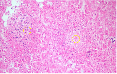 |
Figure 1. Fetal hepatic necrosis |
References:
1. M.L. Anderson. Infectious Causes of Bovine Abortion During Mid- to Late-gestation. Theriogenology 68 (2007), page 474-486
2. NAHMS, Beef 2007-08. Part IV Reference of Beef Cow-calf Management Practices in the United States, 2007-08, page 44
