July 2026
Heinz Bodies and Eccentrocytes: Evidence of Oxidative Injury to the Erythrocytes
By Irene Ulloa Dobles, DVM, Diagnostic Medicine Intern; Co-author: Daniela Hernandez Muguiro, DVM, DACVP (Clinical Pathology)
Erythrocytes perform the critical task of transporting oxygen to all body tissues. Oxygen is a strong oxidant, as it can generate highly reactive derivatives by reacting with iron. This, combined with the fact that erythrocytes are exposed to various chemicals in plasma, makes them particularly susceptible to oxidative damage.1,2 Erythrocytes rely only on a limited arsenal of antioxidant mechanisms to protect against oxidant species, which are constantly being produced. However, when these antioxidant mechanisms are overwhelmed, oxidative damage to the erythrocytes occurs.1,2 But how does this damage occur, and how does it affect these cells?
Mechanisms of Oxidation to the Erythrocytes
Oxidants can damage various components of erythrocytes, each resulting in distinct morphologic changes or features. For example, when oxidants damage the erythrocytes' hemoglobin, it can result in Heinz body formation, whereas damage to the erythrocyte membrane leads to the formation of eccentrocytes.3 There are various mechanisms proposed for how this injury process happens, which are discussed next.
Oxidation of Hemoglobin
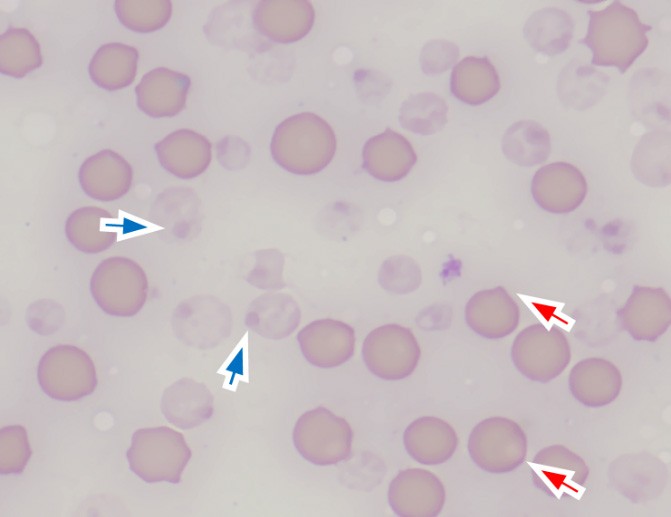
Under normal physiological conditions, a small proportion of oxyhemoglobin is converted to methemoglobin. This process produces hydroxyl radicals, which in turn lead to the formation of reversible and irreversible hemichromes. These hemichromes, also known as denatured hemoglobin, consist of structural changes to the hemoglobin molecule that alter its solubility. Reversible hemichromes can be reduced back to hemoglobin, whereas irreversible hemichromes remain and aggregate. Irreversible hemichromes form when oxidant compounds overwhelm the antioxidant mechanisms of the erythrocytes.2,4 Because these aggregates of irreversible hemichromes are less soluble than normal hemoglobin, they precipitate and form Heinz bodies. Heinz bodies were first described in 1890 by Dr. Robert Heinz as round structures protruding from the erythrocyte surface of humans and animals that had been exposed to certain types of coal tar drugs (Figure 1).1,2,4
 |
|
Figure 1. Intact (red arrows) and ghost erythrocytes (blue arrows) with Heinz bodies. Blood smear of a sheep with copper toxicosis and oxidative hemolytic anemia. Wright’s-Giemsa, 1000x magnification. |
Heinz bodies can also form following oxidation of the sulfhydryl groups of the globin portion of the hemoglobin molecule. Oxidation of the exposed β-93 cysteine residues present in these sulfhydryl groups results in sulfhemoglobin formation. Sulfhemoglobin, like the hemichromes, is less soluble than normal hemoglobin and precipitates to form Heinz bodies that bind to the cell membrane.2,5 Of these two mechanisms described for Heinz body formation, it is believed that hemichrome formation is likely more important, except possibly in cats, which differ from other species in their hemoglobin composition.2
Oxidation of the Erythrocyte Membrane
Membrane structures can also be altered by oxidative injury. Specifically, the sulfhydryl groups in proteins and polyunsaturated lipids in membranes are particularly susceptible to oxidation and lipid peroxidation.3,6 These processes, which produce lipid peroxyl radicals and hydroperoxides, lead to crosslinking and aggregation of membrane and cytoskeleton proteins, resulting in the adhesion of the opposing faces of the erythrocyte membrane. The adhesion of opposing faces of the membrane pushes the hemoglobin inside the erythrocyte to one side, leaving a clear area on the other side, which is known as an eccentrocyte.7,8,9,10
Denatured spectrin dimers and tetramers are believed to be the main components in the crosslinking of the erythrocyte membranes.8 Eccentrocytes have also been thought to form secondary to precipitation of oxidized hemoglobin with bystander oxidation of the membrane.1
Species Variations
Animals’ erythrocytes react differently to oxidation injury; while some species are more prone to the formation of Heinz bodies, others are more prone to eccentrocyte formation. This is because variations in species exist regarding hemoglobin structure and erythrocyte metabolism.1,11 Cat hemoglobin is notable for having 8-10 readily oxidizable sulfhydryl groups, whereas most other species have only two.3,11,12 Cat hemoglobin also readily dissociates from the usual tetrameric form to dimers, which have a greater tendency for auto-oxidation.11 Additionally, a cat's spleen is non-sinusoidal and relatively inefficient at removing Heinz bodies compared to other species. These factors are thought to contribute to the routine presence of low numbers of small Heinz bodies in cat blood (up to 5-10% of erythrocytes).3,11,12
Identifying Heinz Bodies and Eccentrocytes in a Blood Smear
On blood smears stained with routine Romanowsky-type stains, Heinz bodies may appear as eccentric structures at the red cell margin, or they may bulge from the surface of the erythrocytes if large enough (Figure 1b).2 Heinz bodies appear lighter in color or refractive compared to the rest of the hemoglobin in the erythrocytes.5,7,13 They are generally small, around 0.5-1 μm in diameter, although they may be larger. Heinz described them as 1-2 μm in diameter in dogs, rabbits, and guinea pigs. They are largest in cats, where they can be up to a third the diameter of the erythrocytes, although this is usually seen in pathological conditions with increased oxidative injury.5,14 Supravital stains, such as New Methylene Blue (NMB), can be used to highlight and more accurately quantify Heinz bodies, as they may be difficult to identify with routine stains. Staining with NMB highlights Heinz bodies as distinct, round or small granular structures that appear darker blue compared to the rest of the hemoglobin, which makes them easier to identify (Figure 2).5,7,13,14
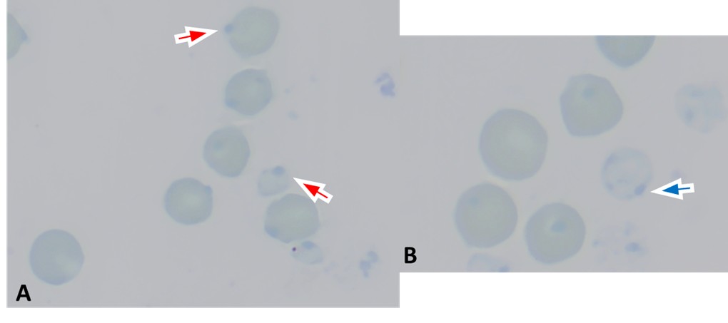 |
| Figure 2. Heinz bodies highlighted with New Methylene Blue (NMB) supravital stain. Blood from the same sheep as Figure 1b. Heinz bodies appear as round structures that are darker blue than the rest of the hemoglobin in intact (A: red arrows, magnified image 1000x magnification) and ghost (B: blue arrow, magnified image from 1000x magnification) erythrocytes. |
Eccentrocytes are erythrocytes characterized by the concentration of hemoglobin on one side of the cell, leaving a clear or pale, hemoglobin-free, blister-like area on the other side. 5,7,9 These are easier to recognize on Romanowsky-type stains compared to Heinz bodies (Figure 3). However, they can also be highlighted with NMB preparations.13
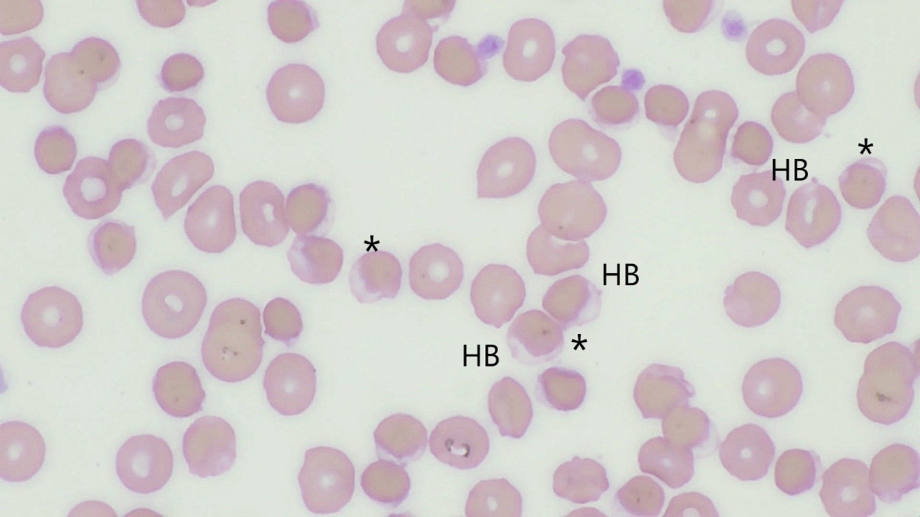 |
| Figure 3. Moderate numbers of eccentrocytes (asterisk), and few small Heinz bodies (HB) are present. Blood smear of a dog with oxidative-hemolytic anemia secondary to Propofol administration (Wright-Giemsa, 1000x, magnification). Note how eccentrocytes have the hemoglobin displaced to one side of the cell, leaving a clear, pale, hemoglobin-free area on the other side. |
Pathogenesis of Oxidative Hemolytic Anemia
Several factors have been implicated in the pathogenesis of oxidative hemolytic anemia, including the mechanical removal of Heinz body-bearing erythrocytes and of eccentrocytes by the spleen (although, as stated above, this appears not to be the case in cats).1,2,4 The oxidative hemolytic anemia has also been attributed to immune-mediated mechanisms. Hemichromes and Heinz bodies have an affinity for membrane protein band 3, making this protein form clusters both on the inside and outside of the erythrocyte membrane. The external clustering of protein band 3 creates a recognition site for autoantibodies to bind to, which leads to their subsequent removal by macrophages.2,4,15Other changes that contribute to hemolysis include cytoskeletal protein crosslinking, cation imbalance, lipid peroxidation, depletion of reduced glutathione, and proteolysis induced by hemichromes.1,2,15
Eccentrocytes also play a role in the development of hemolytic anemia resulting from oxidative damage, as they are more rigid and less able to pass through splenic sinusoids, becoming trapped and subsequently removed by macrophages. They are also more fragile due to damage, an oxidized membrane, and a compromised cytoskeleton, and may spontaneously rupture within blood vessels.
Specific Causes of Oxidative Injury to the Erythrocytes
Several compounds from various sources can cause oxidative damage to erythrocytes. The following are the most reported in domestic animals.
Plants
Members of the Allium family, such as onions, garlic, leeks, and chives, contain aliphatic sulfides, which are oxidative compounds because they decrease glucose-6-phosphate dehydrogenase (G6PD) activity in erythrocytes. This affects the regeneration of reduced glutathione, which is needed to prevent oxidative denaturation of hemoglobin.1,2,4 Most species of domestic animals can be affected by the compounds on these plants; and onion ingestion is the most common cause of Heinz body hemolytic anemia and eccentrocyte formation in dogs.2,7,9 In cats, ingestion of onion soup and baby food containing onion powder has also been shown to produce Heinz body hemolytic anemia.1,7
One of the most severe and possibly deadly causes of Heinz body hemolytic anemia, and eccentrocytes formation in horses, llamas, ponies, and zebras, is caused by ingestion of the plant Acer rubrum, which is red maple leaves, when these are wilted or dried (not fresh). The oxidative compound is thought to be gallic acid, which causes rapid depletion of glutathione.1,2
In ruminants, ingestion of plants in the Brassica family, such as cabbage and kale, may result in Heinz body hemolytic anemia, and the severity of the disease is proportional to the amount of the plant in the diet. The Brassica plants contain a compound that is metabolized by rumen bacteria into the oxidant dimethyl disulfide.1,2
Drugs and Chemicals
In cats, acetaminophen ingestion is likely the most common cause of Heinz body hemolytic anemia. Due to inefficient metabolism of acetaminophen, oxidant metabolites accumulate.1,2,4 As a result, glutathione concentration is decreased and oxidative damage to the erythrocytes occurs. In dogs, acetaminophen ingestion can cause oxidative injury as well, which is characterized by eccentrocytes formation.1,2
Zinc ingestion in dogs, from metal objects such as zinc-containing pennies, nuts, bolts, and other materials containing zinc, like toys and ointments, has been known to cause Heinz body hemolytic anemia. The mechanisms have not been fully elucidated, but they may involve zinc interference with glutathione reductase, or direct damage to the erythrocyte membrane, and protein band 3 clustering.1,2,4 Vitamin K and antagonists are also a reported cause of oxidative injury in dogs. Vitamin K oxidizes hemoglobin and produces an intermediate called semiquinone radical, which contributes to erythrocyte membrane damage. This results in the formation of eccentrocytes and Heinz bodies. 1,2,7 Skunk spray, which contains thiol and other oxidizing agents, can also cause this type of injury to dogs.1,2
In ruminants, copper and selenium deficiency and copper toxicosis have been reported to cause Heinz body hemolytic anemia. The latter is most common in sheep.1,2 Several other chemicals such as propofol, benzocaine, phenothiazine and naphthalene, among others, can also cause Heinz body hemolytic anemia in dogs and cats. 1,2,4,7
Diseases
In specific disease states, Heinz body and eccentrocyte formation is increased and may contribute to anemia. In cats, diabetes mellitus, hyperthyroidism, and lymphoma have been correlated with Heinz body formation. This is most prominent in cats with diabetes. In this disease, oxidative damage is caused by the formation of radicals secondary to glucose auto-oxidation, oxidized plasma lipoproteins, and weakened antioxidant systems. Changes in inflammatory mediators and antioxidant nutrient status also contribute to oxidative damage. In different studies, diabetic cats and dogs that are also ketoacidotic had significantly more Heinz bodies and eccentrocytes, respectively, than nonketotic diabetics.1,2 Therefore, it is thought that ketones may be the main culprits of oxidative injury in cats and dogs with diabetes.
Oxidative injury with eccentrocytes and Heinz bodies formation has been reported in dogs and cats with lymphoma. The exact mechanism of oxidative injury remains unclear. In cats, the magnitude of oxidative injury seems to correlate with the stage of clinical disease, the organs involved, therapy, and the feline leukemia virus status.1,2
Hereditary Membrane Defects And Metabolic Disorders
Erythrocyte membrane transport defects have been described in animals as a potential cause of Heinz body hemolytic anemia when exposed to oxidants, particularly those with defects in amino acid transport involved in glutathione metabolism. This has been reported in Finnish Landrace sheep, thoroughbred horses, and Japanese Shiba and Akita dogs, as an inherited autosomal recessive trait.1,2,7
Hemolytic anemia caused by G6PD deficiency was described in an American Saddlebred colt and a dog, where eccentrocytosis was the primary major morphologic abnormality.1,2,7 Deficiency of Flavin Adenine Dinucleotide (FAD), cofactor of glutathione and cytochrome reductases, in a Spanish mustang mare with eccentrocytosis and methemoglobinemia has also been reported.6
- Behling-Kelly E, Newman A. Anemia Associated with Oxidative Injury. In: Brooks MB, Harr KE, Seeling DM, Wardrop KJ, Weiss DJ, eds. Schalm’s Veterinary Hematology. 7th. Ed. 2022. John Wiley & Sons; 252-259.
- Thrall MA. Regenerative anemia. In Thrall MA, Weiser G, Allison RW, Campbell TW, eds. Veterinary Hematology, Clinical Chemistry and Cytology. 3rd 2022. John Wiley & Sons; 110-138.
- Harvey JW. Erythrocyte biochemistry. In: Brooks MB, Harr KE, Seeling DM, Wardrop KJ, Weiss DJ, eds. Schalm’s Veterinary Hematology. 7th. Ed. 2022. John Wiley & Sons; 166-171.
- Stockham SL, Scott MA. Erythrocytes. In: Stockham SL, Scott MA. Fundamentals of Veterinary Clinical Pathology. 2nd 2008. Blackwell Publishing; 107-222.
- Tvedten H, Moritz A. Reticulocyte and Heinz Body Staining and Enumeration. In: Brooks MB, Harr KE, Seeling DM, Wardrop KJ, Weiss DJ, eds. Schalm’s Veterinary Hematology. 7th 2022. John Wiley & Sons; 181-187.
- Harvey JW, Stockham SL, Scott MA, Johnson PJ, Donald JJ, Chandler CJ. Methemoglobinemia and eccentrocytosis in equine erythrocyte flavin adenine dinucleotide deficiency. Vet Pathol. 2003 No;40(6):632-42.
- Weiss DJ, Tvedten H. Erythrocyte disorders. In: Willard MD, Tvedten H. Small Animal Clinical Diagnosis by Laboratory Methods. 5th ed. 2011. Saunders Elsevier; 38-62.
- Harvey JW. The erythrocyte: physiology, metabolism and biochemical disorders. In: Kaneko J, JW Harvey, Bruss ML, eds. Clinical Biochemistry of Domestic Animals. 5th 1997. Elsevier; 157-194.
- Caldin M, Carli E, Furlanello T, Solano-Gallego L, Tasca S, Patron C, Lubas G. A retrospective study of 60 cases of eccentrocytosis in the dog. Vet Clin Pathol. 2005 Sep;34(3):224-31.
- Brockus CW. Erythrocytes. In: Latimer KS, ed. Duncan & Prasse’s Veterinary Laboratory Medicine: Clinical Pathology. 5th 2011. John Wiley & Sons; 3-44.
- Gibson J, Wadud R, Lu C-Y, Brewin J, Rees D. Oxidative stress and haemolytic anaemia in dogs and cats: a comparative approach. Integr J Vet Biosci. 2019; 3(3): 1–5.
- Schaefer DM. Hematology of cats. In: Brooks MB, Harr KE, Seeling DM, Wardrop KJ, Weiss DJ, eds. Schalm’s Veterinary Hematology. 7th 2022. John Wiley & Sons; 983-992.
- Barger AM. Erythrocyte morphology. In: Brooks MB, Harr KE, Seeling DM, Wardrop KJ, Weiss DJ, eds. Schalm’s Veterinary Hematology. 7th 2022. John Wiley & Sons; 188-197.
- Thrall MA. Erythrocyte production, function and morphology. In Thrall MA, Weiser G, Allison RW, Campbell TW, eds. Veterinary Hematology, Clinical Chemistry and Cytology. 3rd 2022. John Wiley & Sons; 87-99.
- Pires A, Christian JA. Erythrokynectics and erythrocyte destruction. In: Brooks MB, Harr KE, Seeling DM, Wardrop KJ, Weiss DJ, eds. Schalm’s Veterinary Hematology. 7th 2022. John Wiley & Sons; 172-180.