
Cutaneous Habronemiasis in a Horse
By Dr. Giselle Cino
A one-year-old Quarterhorse was presented to the KSUVHC with a two-month history of two large, ulcerated and necrotic cutaneous masses located in the right axillary region and the ventral thorax, respectively. The masses were partially removed 2 weeks prior to presentation and grew rapidly since then. The masses measured approximately 10 cm in diameter and had been treated with antibiotics and topical medications, but never healed.
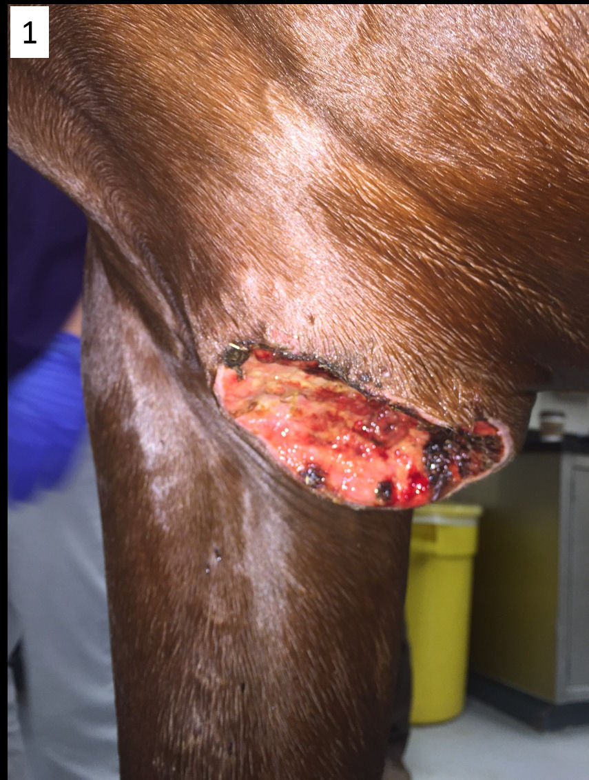 Figure 1 Figure 1
|
Grossly, the masses were raised and ulcerated. The center of the ulcer was necrotic and covered by yellowish-tan granulation tissue. The margins of the ulcer were rounded (Fig.1). On cut sections, the masses were friable, and yellow-white caseous or gritty material was felt through the tissue.
Impression smears from the lesions were submitted for cytopathology and revealed high numbers of eosinophils, along with fewer neutrophils and occasional lymphocytes, consistent with eosinophilic and neutrophilic inflammation.
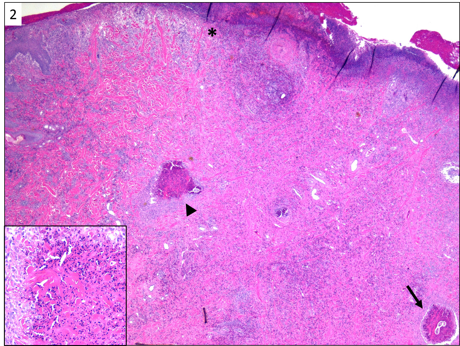 Figure 2 Figure 2
|
Biopsies from both masses were submitted to the KSVDL for microscopic examination. The epidermis over the affected areas is extensively ulcerated and covered by a serocellular crust of fibrin, erythrocytes, eosinophils, and cell debris (Fig 2, asterisk). The remaining epidermis is hyperplastic. The dermis is extensively infiltrated by large numbers of intact and degranulated eosinophils, epithelioid macrophages, plasma cells, and fewer multinucleated giant cells surrounding variable sized flame figures (Fig 2, black arrowhead) and cross sections of nematode larvae (Fig. 2, black arrow). Flame figures are composed of hypereosinophilic collagen fibers (collagenolysis) with cell debris and are occasionally mineralized (Fig 2, insert). Larvae measure ~50 µm in diameter, have an external cuticle with spines (Fig 3, black arrows), coelomyarian musculature, a muscular esophagus, and intestine. Some dead larvae have mineral deposits along the cuticle.
The microscopic lesions are characteristic of cutaneous habronemiasis, a condition caused by the aberrant deposition of larvae of the spirurid nematodes Habronema majus (microstoma), H. muscae, and H. megastoma by transmitting flies. Common sites of habronemiasis in horses include the legs, ventrum, prepuce, urethral process of the penis, and medial canthus of the eyes. Affected horses develop a hypersensitivity response to the larvae characterized by extensive eosinophilic infiltration. Lesions are rapidly progressive and proliferative and consistently pruritic.
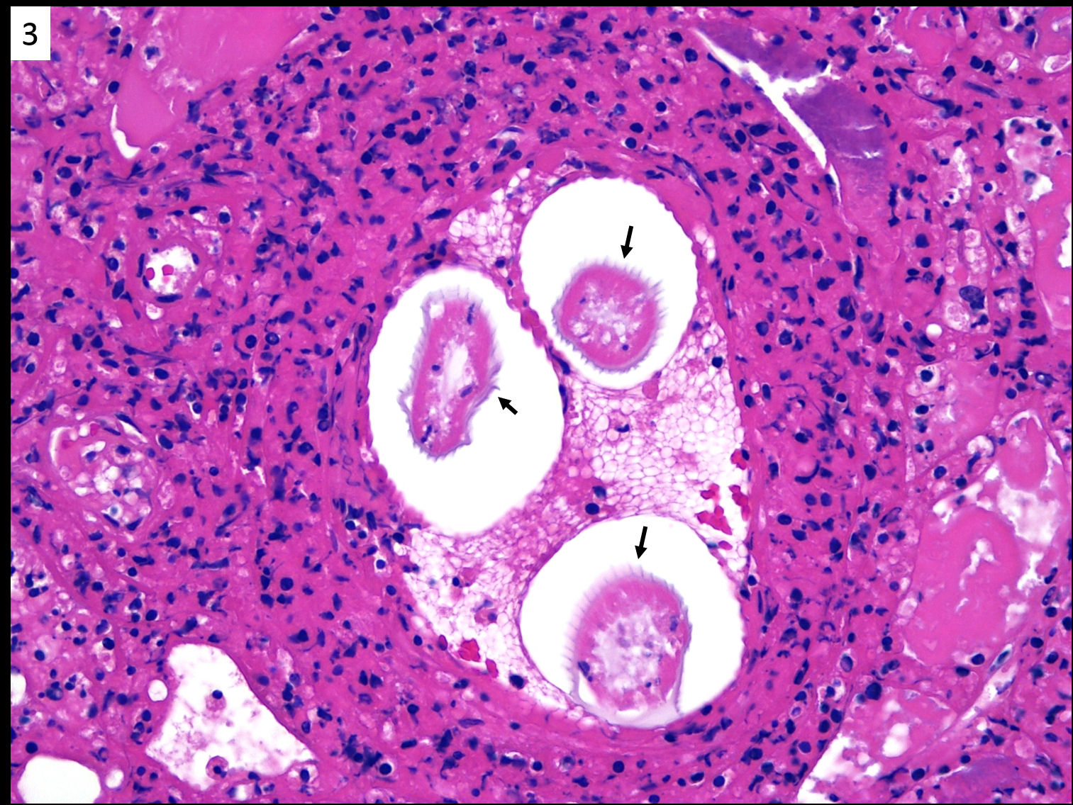 Figure 3 Figure 3
|
Since the gross findings of cutaneous habronemiasis may resemble those of pythiosis, exuberant granulation tissue, botryomycosis, equine sarcoid, and squamous cell carcinoma, the definitive diagnosis requires biopsy and histologic examination of tissue sections by a pathologist.
Next Article: 5th Annual KSVDL Continuing Education Conference
Return to Index