
November 2022
Pinworms in Reptiles
By Jeba R J Jesudoss Chelladurai BVSc, MS, PhD, DACVM
Exotic pet ownership is on the rise and reptiles including bearded dragons, ball pythons, geckos and turtles are being owned by more people than ever before. In this brief, let us look at the diagnosis and management of the most common parasite of reptiles – pinworms (Oxyurid nematodes). The most common pinworm of lizards such as bearded dragons belongs to the genus Pharyngodon, and the most common pinworm of turtles belongs to the genus Tachygonetria. Pinworms are specific to their host species, that is, pinworms of turtles will not infect lizards and vice versa.
Lifecycle
From their infection sites in the large intestines, each female pinworm may lay hundreds of eggs, which are passed in the feces. The eggs need to undergo embryonation outside the host, which happens rapidly in warm temperatures. Infection of the next susceptible host or reinfection of the infected hosts occurs when embryonated eggs are ingested from the environment. Thus, the lifecycle is direct and there is no involvement of an intermediate host.
Pathogenesis and Epidemiology of Infection
Pinworm adults do not usually cause disease in infected reptiles. Some research even suggests that pinworms may support gut health in herbivorous lizards. No clinical signs and/or changes in feces are typically seen. Pinworms in lizards and turtles are commonly seen in captive breeding facilities and when animals are held in extremely poor husbandry conditions. Introducing a new reptile into a tank/vivarium previously occupied by an infected reptile of the same species will cause the introduced animal to become infected.
 |
|
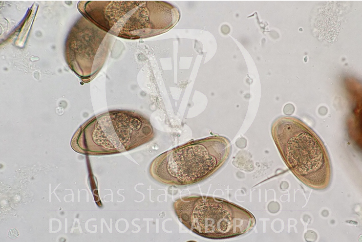
Figure 1: Pharyngodon eggs from a bearded dragon (40x) |
Diagnosis and Treatment
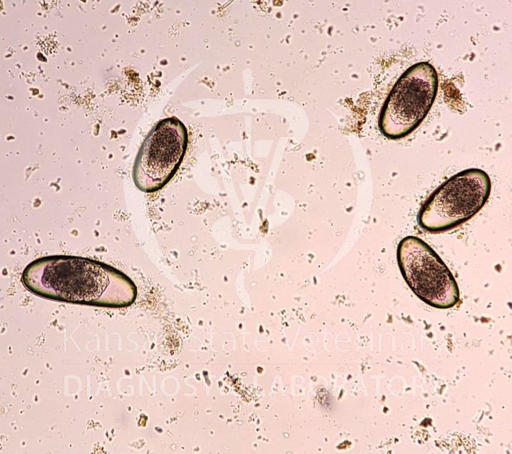
Diagnostic tests can be performed on feces to determine if the reptile is infected. Performing a qualitative fecal float will aid in the microscopic visualization of pinworm eggs. Pinworm eggs of reptiles are yellowish to yellow brown, have a thick egg shell, have one polar operculum and one side of the egg is flattened while the other side is convex. Reports have + (1-10 eggs), ++ (11-50 eggs) and +++ (50 -100 eggs) to denote the number of eggs seen on the slide. Pharyngodon eggs and Tachygonetria eggs are shown in figures 1 and 2. While the eggs may also be visualized on a direct fecal smear, a qualitative fecal float is preferred due to higher sensitivity of the float.
 |
|
Figure 2: Tachygonetria eggs from a turtle (20x) |
Once pinworms are diagnosed in a reptile, fenbendazole may be used for treatment, following the recommendations of the Exotic Animal Formulary (by Dr. Carpenter). An overdose must be avoided since leukopenia may be caused by a toxic dose.
Control and Prevention
Since pinworms eggs accumulate in the environment and may cause reinfection, environmental cleanliness is important in controlling existing infections and preventing reinfections. A plan must be developed by the veterinarian and the owner to eliminate pinworm eggs from the animal’s environment. Relevant information about the animal’s living space, co-housing with other reptiles of the same species/different species, animal’s contact with the outside environment and level of environmental contamination must be considered in making control recommendations.