
July 2018
Thermal Injury: Garden Hose Scalding Syndrome
By Dr. Chanran Ganta
|
|
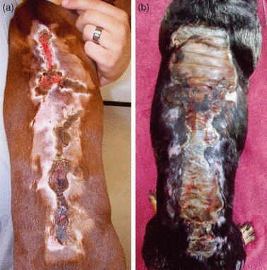
Figure 1. Gross lesions of garden hose scald: Characteristic linear burn pattern along the dorsum, with areas of ulceration and crusting. |
|
|
|
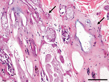
H&E staining: Second degree thermal burn. Note necrotic follicular and adnexal epithelia (arrows). Scale bar= 100 microns. |
Causes of Thermal Injury
Thermal injury in domestic animals is not uncommon; sources include hot liquids, steam, heating pads, hair dryers, drying cages, hot metals such as wood stoves or car engines, fires, friction from rope "scalds," electrical burns from chewing electrical wires, improperly grounded electrocautery units, or lightning strikes. Short term exposure to extremely hot temperatures (e.g. 1 s, 68 °C⁄155°F) and prolonged exposures to lower temperatures (e.g. 5 min, 48.9°C⁄120°F) can produce similar injuries.
Garden Hose Scalding Syndrome
Lesions often manifest on the dorsum of the skin with second or third degree burns and are always linked with a history of the pet being hosed on a hot day with a hose exposed to the sun. The clinical signs of scald injuries usually do not manifest for several days; therefore, the owners may not remember the clinical history. Gross lesions are typical of severe thermal burns that are spread linearly on the dorsum (Figure 1). Changes in the skin include marked erythema, ulceration, edema, and cutaneous necrosis with secondary bacterial infection.
Clinical Features and Diagnostic Criteria
The following 5 criteria are used to diagnose this condition:
1. Clinical history: the dog had been hosed off with water from a garden hose
2. Lesion distribution: linear burn pattern along the dorsum
3. Gross and histological appearance: second or third degree thermal burn
4. Time of year: May to August
5. Geographical location: areas where recorded high temperatures were above 32°C (90°F), such as in the western or southern USA.
Histopathological Features
Specific histopathological features are characterized by "necrosis of hair follicular epithelium with surrounding normal viable dermal collagen" (Figure 2) and are a helpful diagnostic tool in differentiating thermal burns from other similar looking lesions such as toxic epidermal necrolysis, severe erythema multiforme, or insect envenomation. This pattern is the result of the 'tracking' or 'wicking' of heat down the hair shaft like the flame on a candle wick.
Summary
Surgical biopsy of all thermal injuries will help in evaluating the size and depth of the injury, which helps to classify the degree of the burn and its prognosis. All biopsy submissions require adequate clinical history, age of the pet, location of the biopsies following proper biopsy collecting techniques.
For an article on collecting biopsies, please visit:
https://www.ksvdl.org/resources/news/diagnostic_insights/march2016/Biopsies.html
WATCH: "6 Tips for Biopsy Submission", here: https://www.youtube.com/watch?v=79v69x7qLFs
If you have questions about this disease or other diseases or diagnostics, please contact KSVDL Client Care at 866-512-5650 or clientcare@vet.k-state.edu.